With one of the highest death rates worldwide, Cancer leads the pack as one of the most dreadful groups of diseases known to mankind. Cancer is referred to as an unwanted growth of cells in a certain organ or a part of the body. The tumours involved are generally of three types. Benign tumours are not cancerous and cannot spread to different parts of the body. Premalignant tumours are not yet cancerous but may develop cancerous traits later on. On the other hand, Malignant tumours can spread to other organs and parts of the body, making them very fatal to the human body. In recent news from the University of Lincoln, scientists have reproduced a synthetic copy of the Anti-tumour antibiotic in order to combat Drug-resistant bacteria and cancer.
About The Antibiotic – Kedarcidin
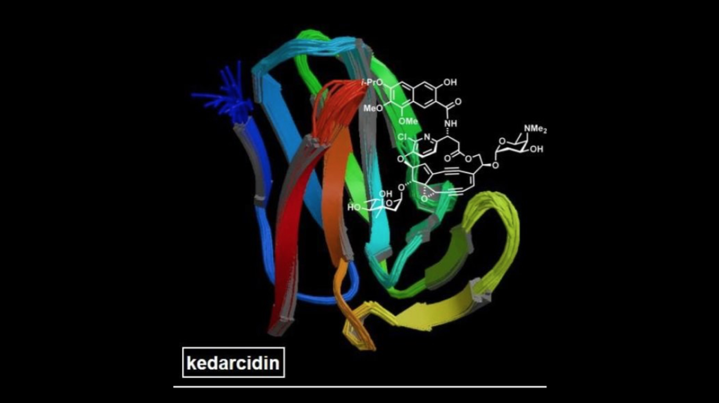
 The antibiotic, or rather, the “super-substance” is called Kedarcidin. It was first discovered almost 30 years ago by a pharmaceutical company in India. Extracted from a soil sample, Kedarcidin’s natural form was unusable as a potential drug to fight cancer. In fact, most antibiotics developed in the last 70 years have been derived out of soil, but in order to use them as a drug for treatments, the antibiotics have to be reproduced via the process of chemical synthesis in the laboratory. Kedarcidin, however, is different from conventional antibiotics.
Mechanism And Structure Of Kedarcidin
 For starters, Kedarcidin is capable of harming tumour cells instead of just killing the bacteria involved (like many other antibiotics). This makes it a very potential candidate as a primary drug in effective cancer treatments. The biological structure of Kedarcidin enables it to harm the DNA structure of the target tumour to a level of complexity that the tumour cannot spread at all. Because of the complex structure of Kedarcidin, scientists were unable to replicate it’s “potential drug” form, until now.
The primary scientists involved in the replication of the aforementioned drug are Dr. Martin Lear (University of Lincoln, UK) and Professor Masahiro Hirama (Tohoku University, Japan). As per the scientists, the molecular structure of the antibiotic drug resembles an egg of sorts. Moreover, Kedarcidin has a reactive core which is sheathed by a layer of protein. As per reports, almost 10 years were spent in the deduction of the molecular integrity of the antibiotic.
 Also Read: You Can Now 3D Print Full Human Organs Including A Live Working Heart
It is estimated that the number of cancer cases per year worldwide will rise to 23.6 million by the year 2030. Considering the latest advancements in the “cancer-fighting” drug, the numbers may experience a steep drop, and for the better. Aggressively tackling the tumours with Kedarcidin may help scientists learn a lot more about the techniques which the antibiotic uses to counter cancerous cells in leukaemia and melanoma, for instance.

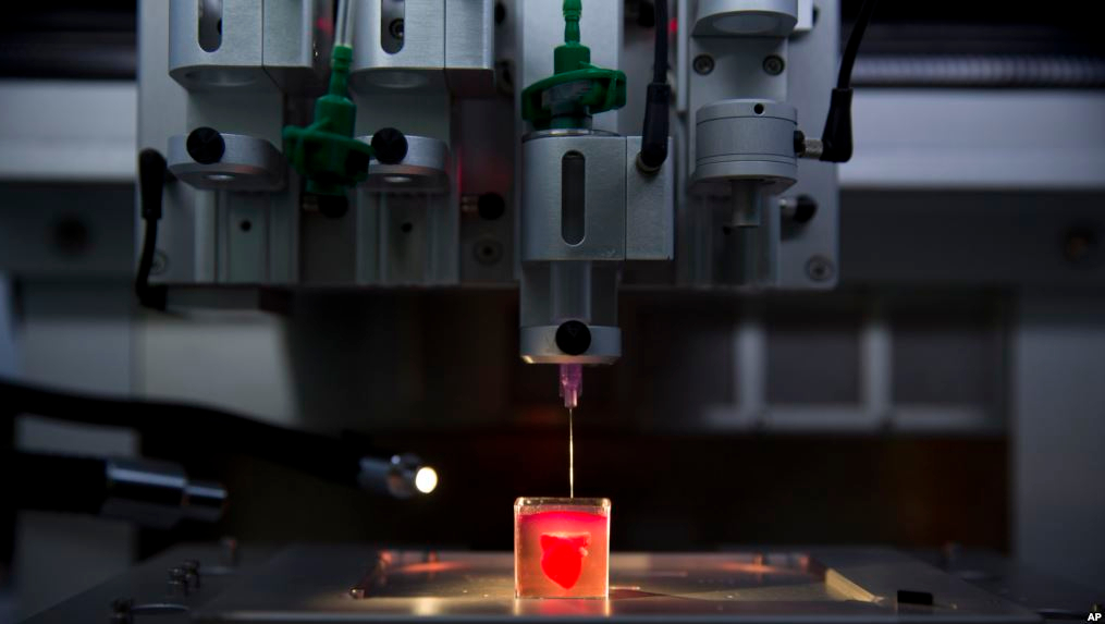 Using a patient’s own cells, the researchers from Tel Aviv University created the heart on a high resolution 3D printer. The university has announced their success story while showcasing the printed heart. The printing experiment was deduced as one of the most major medical breakthroughs to date. The research findings were published in the Journal of Advanced Science. The team involved in the experiment was led by university professor Tal Dvir.
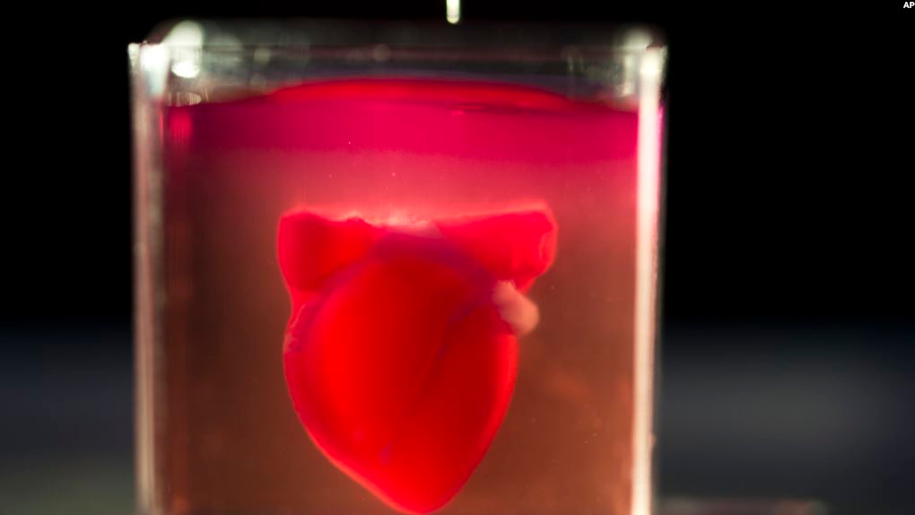 As per Tal Dvir, this was actually the first time that human cells were incorporated with 3D printing technology to formulate a complete replica of a heart. The 3D printed heart is design to perform in the real world, so it comprises of blood vessels which help the heart to pump blood. Previously, only non-vessel tissues could be printed using the combination of medical methods and technology, but a new technique adopted by the researchers made it possible to design a life-like 3D-printed heart. Fatty tissue from patients was used as the “fuel” or “ink” of the 3D printer.